Revised 2011
OBJECTIVES:
-
To gain experience in basic plant pathology laboratory techniques.
- To determine whether local plants contain active ingredients against bacterial organisms.
INTRODUCTION:
Although there are many methods used against bacterial diseases, bacteria are usually very difficult to control (1). Frequently, a combination of several control measures is required to combat a given bacterial disease. Soil infested with plant pathogenic bacteria can be sterilized with steam or dry heat, or with certain chemicals. These techniques, however, are practical only in greenhouses and in small beds or frames. The use of chemicals has been much less successful for the control of bacteria than for the control of fungi. Copper compounds produce the best results against bacteria, but they seldom provide satisfactory control of the disease when environmental conditions favor development and spread of the bacteria. Bacterial strains resistant to copper fungicides are quite common. Copper compounds can also be phytotoxic on certain plant species. Antibiotics have been used against certain bacterial diseases with mixed results. Treatment of plants harboring bacteria has been successful under experimental conditions, but the application of antibiotics has been less successful in practice. Antibiotics are also expensive, and the antibiotics valuable for human therapy are not allowed to be used in agriculture (4).
One bacterial disease of plants, called crown gall, is caused by tumorigenic strains of the Gram-negative bacterium
Agrobacterium tumefaciens. The bacterium overwinters in infested soils, where it can live as a saprophyte for several years. When host plants are growing in infested soils, the bacterium enters the roots or stems near the ground through wounds caused by factors such as freeze damage, grafting or mechanical injury. The bacterium finds plants by detecting phenolic substances produced by wounded plant cells (1). Once inside the plant tissue, the bacterium moves from cell to cell, stimulating surrounding host cells to divide at a rapid rate. The bacterium does this by transferring a piece of its own genetic information, or DNA, into the plant cell. This piece of genetic information does not come from the chromosome of the bacterium, but from a separate piece of DNA called a plasmid (3). The
A. tumefaciens plasmid is called the tumor-inducing or Ti-plasmid, and the piece of DNA that is transferred to the plant is called the T-DNA. Following transfer to the plant, the T-DNA becomes integrated into the chromosomes of the plant cell (3). Genes on the T-DNA cause the plant cell to divide repeatedly, forming the gall or mass of undifferentiated tissue, and to produce chemicals called opines, which are used by the bacterium as food. The bacterium itself lives and multiplies in the intercellular spaces of the gall (3). Young, soft galls are easily injured and attacked by insects and saprophytic microbes, which cause the outside cell layers to decay and discolor. The breakdown of the gall releases bacteria back into the soil, and the bacteria are free to infect new plants (1).
Symptoms of
crown gall are small galls on roots and at the crown of woody plants and some field crops (2). Affected plants may become stunted, produce small, chlorotic leaves, and are more susceptible to adverse environmental conditions, especially winter injury (1). Severely infected plants may die.
Plants representing over 93 plant families are susceptible to crown gall (2). There is no cure for the disease; preventive measures and resistance breeding are the methods now used against
A. tumefaciens (L. Kovacs, personal communication). Some strains of
A. tumefaciens are sensitive to an antibiotic produced by
A. radiobacter, a closely related soilborne bacterium that does not infect plants (2). In some cases, biological control of the disease can be gained by soaking seeds, seedlings or rootstocks in a suspension of
A. radiobacter. Unfortunately, some strains of
A. tumefaciens are able to develop resistance to the antibiotic produced by
A. radiobacter (1).
In this lab, students will simulate tests done to determine whether plants or other substances contain active ingredients against bacterial organisms. Test materials may include local plants and dried herbs known for their ability to control plant pathogens. Also, while there are few substances known to inhibit bacterial growth, there are many substances known to inhibit fungal growth. It is possible that some of those fungal inhibitors could be effective in the control of bacterial infections. The following chemicals and products, many derived from plants, have been effective in inhibiting growth of various pathogenic fungi: sulfur, baking soda, mineral oil, baking soda-mineral oil combination, and
Rumex sp. (dock) (8);
Azadirachta indica (neem oil), jojoba oil, cinnamon oil, soybean oil, compost tea,
Equisetum arvense (horsetail plant) and chlorite mica clay (6);
Reynoutria sachalinensis (giant knotweed) and garlic (9). Other ingredients listed on various labels of 'natural' fungicides include: yeast, tea tree oil, citric acid, mint oil, onion exudates, calcium, weak chamomile tea, cloves, black walnut hull powder, golden seal powder, cayenne and calendula. Active ingredients can include antiseptics, astringents, antibiotics and toxins.
Students will collect samples of plants that might inhibit growth of
A. tumefaciens. Solutions containing extracts of the plants will be tested for their ability to inhibit bacterial growth by soaking carrot disks in the solutions and then inoculating the disks with
A. tumefaciens.

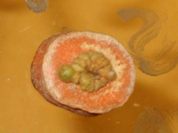
Figure 1. Gall formation on a carrot slice inoculated with
A. tumefaciens. Click image for a more detailed view. |
CLICK HERE FOR INSTRUCTOR'S NOTES
MATERIALS:
Inoculum of
Agrobacterium tumefaciens
Carrots
Plant foliage
Alcohol (95% ethanol)
Bleach (10% solution)
Distilled water (sterile)
Petri dishes
Filter paper (sterile)
Parafilm or Scotch tape
Permanent markers
Mortars and pestles (sterile)
Pipettes (sterile)
Forceps (sterile)
Inoculating loop (sterile)
PROCEDURES:
- Wipe all work areas with disinfestant such as Lysol or a 10% bleach (Clorox) solution. Wash hands with soap and water.
- Obtain one petri plate and three carrot disks for each type of material you wish to screen (maximum of three per student). Students may work separately or in pairs.
- Prepare petri plates by labeling them with student names, date, and the name of the material being tested for antibiotic properties. Be careful to keep the plates covered to prevent contamination.
- Using sterile forceps, place a sterile piece of filter paper on the bottom of each plate.
- Surface sterilize plant material by soaking it for 10 minutes in a 10% commercial bleach (Clorox) solution.
- Rinse plant material with sterile distilled water two times to remove residual bleach from the surface.
- Using a sterile mortar and pestle, grind up around 2 g of plant material with the addition of around 10 ml sterile distilled water. Add more sterile distilled water, a few drops at a time, if necessary to obtain a solution containing the plant extract.
- Sterilize the mortar and pestle after use with an autoclave or by soaking them for 10 minutes in a 10% bleach solution.
- Repeat steps 4 through 8 for each substance being screened.
- Transfer the liquid from the mortar into the labeled petri dish. (Filter if necessary so solid plant materials are not transferred to the dish.) Do this step as quickly as possible so the plate will be open to the atmosphere for as short a time as possible, but not so quickly that careless mistakes are made.
- Slice carrots that have been surface sterilized and rinsed in sterile distilled water into disks about 1 cm thick. Make a small notch on the basal side of each disk to distinguish the basal side from the apical side.
- Using sterile forceps, place three carrot disks in each petri plate that contains an extract you prepared from the ground plant material. After one minute, turn the disks over. Again, keep the plates covered as much as possible to prevent contamination. After soaking the disks, discard (pour out) any plant extract left in the plate.
- Using a sterile inoculating loop, carefully transfer a small amount of
Agrobacterium tumefaciens inoculum to the center of the basal side of each carrot disk.
- In a separate petri plate, saturate each side of three carrot disks with sterile distilled water (control). Inoculate with
A. tumefaciens as in step 13.
- Seal all petri plates with Parafilm or Scotch tape.
- Incubate at room temperature, 20 to 25.5 C (68 to 78 F).
- Examine the carrot disks daily, looking for gall formation.
- Record any results during the next three or four weeks.
OBSERVATIONS:
Identify materials that were screened by students in this laboratory exercise, and rate their effectiveness as inhibitors of crown gall in the following table.
List of Plant Materials |
Degree of Sensitivity Rating |
| | |
| | |
| | |
| | |
| | |
| | |
| | |
| | |
| | |
| | |
| | |
| | |
| | |
| | |
Rating of degree of sensitivity
0 = no effect (gall formation on most or all of the disk),
1 = slightly effective (more than 1/3 of the disk is covered with gall),
2 = effective (less than 1/3 of the disk is covered with gall),
3 = highly effective (no gall formation) |
CONCLUSIONS AND QUESTIONS
- Which plant extract(s) inhibited growth of galls? Do these substances have any known similarities?
- If inhibition occurred, was it slightly effective, effective, or highly effective?
- If inhibition was slightly effective, how might the effectiveness be improved?
- Which of the plant materials or other substances had no effect on the growth of galls? Do these substances have any known similarities?
- Why was it important to use sterile techniques in this investigation?
- What factors could have affected the effectiveness of the screened substances in this investigation?
- Why do plants vary so much in their active ingredients? What are the adaptive features of these active ingredients?
- How might the knowledge gained from this lab exercise be used in agriculture to control
Agrobacterium tumefaciens?
LITERATURE CITED:
- Agrios, G.N. 1997. Plant pathology. 4th ed. Academic Press, San Diego, CA.
- Kado, C.I. 2002. Crown gall. The Plant Health Instructor. DOI:10.1094/PHI-I-2002-1118-01
http://www.apsnet.org/edcenter/intropp/lessons/prokaryotes/Pages/CrownGall.aspx
-
D'Arcy, C.J. and D.M. Eastburn. 2003. Crown gall. Plants, pathogens and people website.
http://www.ppp.uiuc.edu
- Deacon, J. 2002. The Microbial World: Biology and control of crown gall. Institute of Cell and Molecular Biology, and Biology Teaching Organisation, Univ. of Edinburgh.
- Hauser, J.T. 1986. Techniques for studying bacteria and fungi. Microbiology Dept., Carolina Biological Supply Co., Burlington, NC.
- Kuepper, G. and R. Thomas. 2002. Powdery mildew control in cucurbits. Current topics, Appropriate Technology Transfer for Rural Areas (ATTRA), Fayetteville, AR.
- McManus, P. S., and Stockwell, V.O. 2001. Antibiotic use for plant disease management in the United States. Online. Plant Health Progress doi:10.1094/PHP-2001-0327-01-RV.
http://www.plantmanagementnetwork.org/pub/php/review/antibiotic/
- Peet, M. 2001. Sustainable practices for vegetable production in the South. North Carolina State Univ.
- Quarles, W. 2002. Least-Toxic Controls of Plant Diseases. Brooklyn Botanical Garden, Brooklyn, NY.
