PURPOSE AND BACKGROUND INFORMATION
Background
The discipline of Plant Pathology encompasses the study of a diverse range of entities: fungi, bacteria, protozoa, viroids, and viruses. One of the smallest and most challenging to teach about are viruses. Plant viruses are submicroscopic, parasitic and sometimes pathogenic agents composed of nucleic acid; most often surrounded by a protein coat [Crawford, 2018; Cann, 2011]. Viral plant pathogens are a large (more than 1800 species) and highly diverse group. Unlike many other plant pathogens, viruses are too small for learners to observe with their eyes or even with a light microscope. They are also quite complex in structure and function despite their limited size and coding capacity. Therefore, to teach and learn about viruses, both instructors and learners have to employ abstract reasoning.
Abstract reasoning is the ability to analyze information, detect relationships and patterns, as well as solve problems on a complex and impalpable level. The discipline of virology offers an excellent opportunity for the intrinsic integration of abstract reasoning and knowledge at multiple levels: molecular, cellular, and organismal. Our abstract reasoning ability has been shown to be highly correlated with our working memory capacity [Capon et al., 2003; Kyllonen and Christal, 1990; Markovits et al., 2002]. Working memory, also known as short-term memory, is a limited capacity cognitive system for storing and managing information required to process and carry out complex tasks such as learning, reasoning, and comprehension. Working capacity varies among individuals, which translates to variations in aptitude for abstract reasoning among learners [Evans, 2003; Oberauer et al., 2007]. In other words, how much and what type of information is retained in our short-term memory, differs based on each individual's experiences, interests, and perspective. This in turn impacts their aptitude or potential to reason abstractly or visualize things they cannot see and is especially important considering that many learners come into this required class with highly negative opinions about viruses. These range from a fear of viruses as pathogens to feeling intimidated about the molecular aspects, perceptions which negatively impact the type of information learners retain and can lead to inhibition or distortion of their reasoning of abstract concepts and ultimately can be counterproductive to learning.
So how do we facilitate understanding of abstract concepts such as a virus, a mono-/multi-partite genome and/or virus particle in a university level science classroom setting where the capacity of each learner for abstract reasoning may vary significantly? One possible answer would be to use a variety of teaching techniques that motivate and engage learners in the learning process. This is especially important with online teaching being such an integral part of the educational matrix. Studies have found that using active learning strategies like project-oriented courses and cooperative learning formats enhance learners' ability to find information on their own, and to become better problem solvers [Goodwin et al., 1991; Miller and Groccia, 1997].
We found that our 'Build Your Own Virus Particle' exercise accomplished more than was initially intended. It built the confidence of learners intimidated by virology literature, who through active participation, realized they were able to read and learn from peer-reviewed journal papers containing research on plant viruses. A change in the attitudes, opinions and thought process of some learners who viewed viruses with fear was also noted. We observed that implementation of our active learning strategies early in the course improved learners understanding of other abstract concepts (such as viral replication and protein expression) that were taught later in the course. When asked to describe the best aspects of the experience, the majority of learners used 'fun' and 'creative' as descriptors. This exercise also provided an opportunity for the instructor to work one-on-one with learners which increased learning and is a positive aspect of any course.
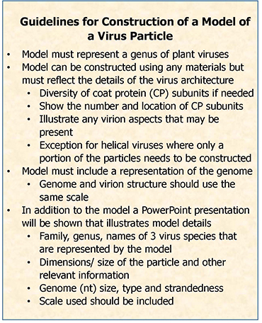
 Figure 1. Virus model construction guidelines provided to learners. Figure 1. Virus model construction guidelines provided to learners.
|
Our 'Build Your Own Virus Particle' exercise encompasses many essential aspects of a graduate scholar experience. Initiating the course/project with a lecture is familiar to learners and allows for a slow progression into what some may refer to as a role reversal. For the duration of the project, instructors take a backseat role and become facilitators. In other words, instructors don't hold all the answers, but rather enable learners to find appropriate resources as well as help them navigate their critical thinking process. This gives learners both the opportunity and responsibility to go search for answers and share these with their peers. This guarantees their engagement with the activity and encourages discussion within and between groups as well as between individuals. It also helped learners develop a number of useful skills: research, cooperation, delegation, communication and teaching. Active learning activities such as the 'Build Your Own Virus Particle' offer learners a hands-on approach to improve understanding of abstract concepts.
MATERIALS AND METHODS
Little preparation for the exercise is required by the instructor. Learners can use any materials that illustrate the type of virus structure they have selected and allow them to meet the Guidelines (Fig.1) and Grading Rubric (Table 1). Their choice of materials or software is unrestricted, and learners are encouraged to be creative. Learners have used cloth, Styrofoam, cotton balls, toothpicks, cardboard and even 3-D printing in the past. The cost to the learners has been minimal, ranging from $20 to $200 (for 3-D printing), but generally learners spend under $50. Learners will also need to be able to use Microsoft PowerPoint to present an explanation of the model to the class and have access to a library with virology journals.
Table 1. Project evaluation rubric used to grade virus models and presentations.
|
ITEM |
POINTS |
PTS EARNED |
| Model correctly represents a genus of plant viruses | 15
|
|
| At least 3 species are listed that possess similar architecture | 5
|
|
| Model is to scale, and scale is presented in the MS PowerPoint | 10
| |
Model illustrates diversity of coat protein subunits if there is variation
| 5
| |
| Includes correct number of coat protein subunits for particle | 10
| |
Location of coat protein subunits is shown accurately on the model
| 5
| |
| Genome representation included in or with model | 10
| |
| Length of genome is to scale | 5
| |
| Correct nomenclature/orthography was used in MS PowerPoint | 3
| |
| Visual clarity of MS PowerPoint | 2
| |
|
70
|
|
OUTLINE FOR INSTRUCTORS
Project Design/Logistics

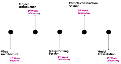
Figure 2. Timeline of the 'Build Your Own Virus Particle' exercise. |
Description: Our 'Build Your Own Virus Particle' exercise requires that learners, either individually, or in small teams, construct a model of a virus structure and deliver a presentation explaining the details of the structure and details of their model. This could be accomplished in as little as four weeks, beginning with a lecture on virus architecture, followed by three class meetings designed to 1) allow learners to express concerns about their project and have questions answered, 2) provide learners time and space to build their virus particle, and 3) allow for learners' presentations of their models.
Timeline: The project timeline is depicted in Fig. 2 and starts with a classroom lecture on virus architecture during the first week of the term. The project goals, guidelines and evaluation rubric are given to the learners with the assignment. Individuals or teams (two or three learners, formed by random selection) are assigned one of five types of virus structures (helix, bacilliform, geminate, icosahedral or enveloped) at random by the drawing of tiles from a beaker (Fig. 3). They are then given a week to research, discuss and formulate a plan using the information from the lecture, appropriate websites, as well as appropriate journal articles that they must find. Part of this research requires that the learner(s) identify a virus with the structure that they have been assigned to model. This is often a moment that generates questions from the learners and allows them to directly interact with the instructor.
A brainstorming session is set during the second week of the course (project mid-point) to help learners follow the guidelines and to answer questions that they may have. Some guidance on the choice of virus for the model is often needed. But we have found these interactions to be highly valuable for establishing rapport, clearing up preconceived misconceptions and establishing confidence if not enthusiasm in the learners.

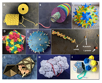
Figure 4. Examples of learner-constructed models of virus architectures illustrating the diversity used to meet the guidelines. Two models of a helical virus structure, in each case the learners modelled a tobamovirus, and both models illustrated the correct number of coat protein subunits per rotation.
A. Model constructed of foam, metal spring and yarn and
B. Model carved from Styrofoam with the RNA genome represented by a pipe cleaner that resembled a published formation of the ssRNA.
C-D. Two models of T=3 icosahedral structure.
C. Model of the exterior of a tombusvirus model using colored paper to indicate the three variations of the coat protein.
D. Model is that of the exterior of a cucumovirus which was constructed of colored cardboard and clay. The 3-dimensional representations of the coat protein subunits of a hexamer and pentamer was created in clay. E: Model of the exterior of an enveloped pleomorphic architecture using an orthotospovirus as the model. Model was constructed of Styrofoam, so not very representative of a pleomorphic shape, and two types of metal pins indicating the 2 glycoproteins in the surface of the envelope.
F. Representation of the L RNA of an orthotospovirus showing the attachment of proteins to the ssRNA genome. The “ssRNA" was wrapped around a pencil to indicate the helical nature of the genome component.
G. Model of non-enveloped bacilliform alfamovirus) composed of cardboard and paper with images of the coat protein subunits, and pipe cleaners representing the ssRNA of the RNA 1 component of this tripartite virus.
H-I. Two models of geminate architecture of a mastrevirus.
H. This model was printed using a powder bed and ink jet head 3D printer. The ribbon represents the ssDNA genome and was pulled out to be visible. The learners obtained the .OBJ file from M. Agbandje-McKenna (Univ. of Florida) who generated the model using cryo-electron microscopy.
I. This model was constructed of cardboard, paper, and used two each side of the particle to show the arrangement of the coat protein subunits using ink on one side and clay polymer on the other.
|
In the third week of the course, learners are given some time during class or lab to start building their virus particles. This provides an opportunity for last minute questions (like those related to the accurate representation and scaling of their assigned viral structure) to be brought up and insures that each learner actively participates in constructing their model. A week later, each learner or team presents its model to the class and explains the details and what each of the materials on the model represents with the help of a Microsoft PowerPoint presentation. The presentation is followed by a brief session answering the instructor's and classmates' questions. If the class is in-person, then learners are given time to view and handle all the models at the end of the session.
Examples of models made by learners for this class exercise are shown in Fig. 4. These images illustrate some of the diverse materials that learners have used. It is important to mention that while learners have to model an entire icosahedral structure, viruses with helical structures, due to their extreme length, are allowed to be presented as partial (not full length) models.
Grading Rubric: Learners are given the evaluation rubric (Table 1) with the assignment to help guide their virus particle construction. Models are judged based on accuracy and inclusion of assigned structure details, and learners should demonstrate originality and greater understanding of their chosen virus architecture, and not on visual attractiveness or difficulty of the materials used. In grading we made allowances for the differences in the amount of information available on the different structures. Pleomorphic structure proved challenging for almost all learners that were given this model. We did observe that in general the more information that was available the greater the detail in learners' models. The grade point value of this exercise was nearly equivalent to an exam (70 points). This was done to increase motivation in the learners and to allow them to allocate the time needed to make the model a successful learning tool. In practice, grades on these models typically ranged from the lower 60s to the maximum of 70. The 'Build Your Own Virus Particle' exercise was part of a low risk assessment approach which included weekly quizzes, a mid-term and comprehensive final exam and a case study of a virus species in the form of a narrated PowerPoint presentation.
Supplemental Information
1. Hulo, C., De Castro, E., Masson, P., Bougueleret, L., Bairoch, A., Xenarios, I., Le Mercier, P. 2011. ViralZone: a knowledge resource to understand virus diversity.
Nucleic Acids Research, 39 (Database issue): D576-82.
https://viralzone.expasy.org/
2. ICTV Virus Profiles (Microbiology Society).
https://www.microbiologyresearch.org/content/ictv-virus-taxonomy-profiles
Literature Cited:
1. Berman, H.M., Westbrook, J., Feng, Z., Gilliland, G., Bhat, T.N., Weissig, H., Shindyalov, I.N. and Bourne, P.E. 2000. The Protein Data Bank. Nucleic Acids Research, 28: 235-242. doi:10.1093/nar/28.1.235, https://pdb101.rcsb.org/learn/paper-models
2. Cann, A.J. 2011. Principles of Molecular Virology (5th Edition). Academic Press, London, UK.
3. Capon, A., Handley, S. and Dennis, I. 2003. Working memory and reasoning: An individual differences perspective. Thinking & Reasoning. 9:203-244.
4. Crawford, D.H. 2018. Viruses: A very short introduction. Oxford University Press, New York, USA.
5. Evans, J.S.B. 2003. In two minds: dual-process accounts of reasoning. Trends in Cognitive Sciences, 7:454-459.
6. Goodwin, L., Miller, J.E. and Cheetham, R.D. 1991. Teaching Freshmen to Think: Does Active Learning Work? BioScience, 41:719-722.
7. Kyllonen, P.C. and Christal, R.E. 1990. Reasoning ability is (little more than) working-memory capacity? Intelligence, 14:389-433.
8. Markovits, H., Doyon, C. and Simoneau, M. 2002. Individual differences in working memory and conditional reasoning with concrete and abstract content. Thinking & Reasoning, 8:97-107.
9. Miller, J.E. and Groccia, J.E. 1997. Are four heads better than one? A comparison of cooperative and traditional teaching formats in an introductory biology course. Innovative Higher Education, 21:253-273.
10. Oberauer, K., Süß, H.M., Wilhelm, O. and Sander, N. 2007. Individual differences in working memory capacity and reasoning ability. Pages 49-75 in Conway, A., Jarrold, C. and Miyake, A., (Eds.). Variation in working memory (p. 49–75). Oxford University Press.
11. World of Viruses: Curricula and Reviews. Retrieved July 6, 2020 from http://worldofviruses.unl.edu/category/curricula/?activity=Models
